Virus causing serious mortalities in Pacific white shrimp in China

A newly discovered iridescent virus that causes severe disease and high mortality in farmed Pacific white shrimp (Litopenaeus vannamei) in Zhejiang, China, has been verified and provisionally named as shrimp hemocyte iridescent virus (SHIV). This article summarizes the original publication (DOI:10.1038/s41598-017-10738-8).
Editor note: There are 12 co-authors in this study but we only list the corresponding one. Please consult the original publication for names and affiliations of all co-authors.
Background
In recent years, epizooties (localized outbreaks) of infectious diseases in farmed L. vannamei have caused massive economic losses in China. SHIV was first detected and identified in samples of pond-farmed L. vannamei (2 to 3 cm) collected from a farm in Zhejiang Province, China, in December 2014. Affected animals experienced massive die-offs and exhibited hepatopancreatic atrophy with fading color, empty stomach and guts and soft shells. Results of testing with polymerase chain reaction (PCR) techniques showed that samples were negative for various known shrimp viruses, including WSSV, IHHNV, VPAHPND, YHV and TSV11.
Also, results of an epidemiological survey indicated that this might not be the first outbreak at this farm. A total of 89 out of 575 L. vannamei individuals, 5 out of 33 Chinese white shrimp (Fenneropenaeus chinensis) individuals, and 5 out of 10 giant freshwater prawns (Macrobrachium rosenbergii) individuals tested SHIV-positive in samples collected during 2014 to 2016 in 20 counties of five provinces over China, raising the concern that the virus may have widely spread to surrounding shrimp farming areas.
Jie, SHIV, Table 1
| Shrimp species | Negative | Positive | Total | Positive rate |
|---|---|---|---|---|
| L. vannamei | 486 | 89 | 575 | 15.5% |
| F. chinensis | 28 | 5 | 33 | 15.2% |
| Mb. rosenbergii | 5 | 5 | 10 | 50.0% |
| Mp. japonicas | 7 | 0 | 7 | 0.0% |
| TOTAL | 526 | 99 | 625 | 15.8% |
The clinical symptoms of SHIV infection include slight loss of color on the surface and cut of hepatopancreas, empty stomach and guts, soft shell in partially infected shrimp and slightly reddish body in one-third of individuals. These symptoms are not similar to those caused by the infection with other alleged iridovirus in other species of penaeid shrimp, sergestid shrimp or freshwater lobsters.
Histological sections revealed that the disease might be caused by a suspected viral infection with obvious pathogenic symptoms, which did not match the histopathologic characteristics of infection with any known viruses or acute hepatopancreatic necrosis disease (AHPND). Using high-throughput DNA sequencing technologies, we determined that a potential iridescent virus was present in one sample, and that the phylogenetic analysis of the two proteins did not support this iridescent virus – provisionally named as shrimp hemocyte iridescent virus (SHIV) – as belonging to any known genus of family Iridoviridae.
Challenge tests and histopathological examination
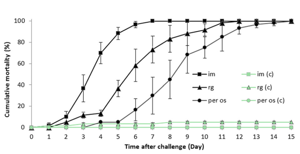
We conducted challenge tests via invasive intramuscular injections or non-invasive mode including reverse gavage or per os infection that could successfully pass a filterable infectious agent from the diseased L. vannamei to healthy animals, and caused similar gross clinical symptoms and pathological changes, proving that this virus is the causative agent of the disease.

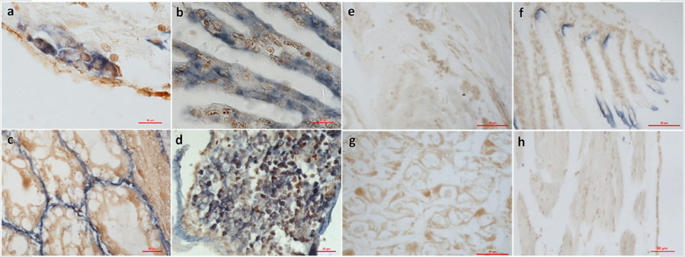
We also we used In Situ Hybridization (ISH) with a specific probe to detect SHIV in sections of infected L. vannamei. In situ hybridization (ISH) is a molecular technique that shows the location of specific nucleic acid sequences in tissues or on chromosomes – this is a critical stage to understand the regulation, organization and function of genes.
The probe reacted to the histological lesions in hematopoietic tissue and hemocytes in gills, hepatopancreas and periopods obtained from a sample, as well as in tissues from the challenge experiment described in this study. Our results fulfilled Rivers’ postulates – created in 1973 by Thomas M. Rivers to establish the role of a specific virus as the cause of a specific disease – for the demonstration of a viral etiology.

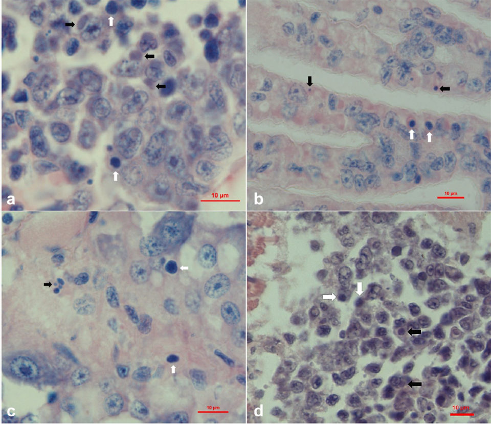
Histopathological examination of sample tissues revealed basophilic inclusions and pyknosis in hematopoietic tissue and hemocytes in gills, hepatopancreas, periopods and muscle. Using the technique of viral metagenomics sequencing (from genetic material recovered directly from environmental samples), we obtained partial sequences we interpreted as potential iridoviridae. And phylogenetic analyses using amino acid sequences of major proteins revealed that it is a new iridescent virus but does not belong to the five known genera of Iridoviridae.

After having compared the morphological, physiological and phylogenetic characteristics of our samples with those of other iridescent viruses, we temporarily assign the etiological agent as shrimp hemocyte iridescent virus (SHIV), which could cause shrimp hemocyte iridescent virus disease (SHIVD). We suggest a new genus of Iridoviridae, Xiairidovirus, which means shrimp iridescent virus.
Perspectives
Through isolation, reinfection and histopathological characterization, we have revealed that SHIV is a new virus in family Iridoviridae and a pathogen of L. vannamei. We have additionally developed an ISH assay and a nested PCR method for the specific detection of SHIV.
The findings of our study emphasize the need for professionals well prepared in aquatic animal health and experienced farmers in the shrimp aquaculture industry to pay closer attention to SHIV and to take more effective measures for preventing the disease outbreaks and economic losses caused by SHIV.
Author
-
Dr. Jie Huang
Corresponding author
Yellow Sea Fisheries Research Institute
Chinese Academy of Fishery Sciences
Qingdao, China
Tagged With
Related Posts

Intelligence
GOAL 2018: A call for collaboration
From GOAL 2018: A global effort to share timely and accurate production data is what the aquaculture industry needs to thrive in today’s ever-changing marketplace.

Health & Welfare
Clinical case report: EMS/AHPND outbreak in Latin America
Behind the successful control of Early Mortality Syndrome (EMS), or Acute Hepatopancreatic Necrosis Disease (AHPND), in a Latin American shrimp nursery.

Health & Welfare
New procedure controls TSV in white shrimp
The authors have established a procedure to reduce the impacts of Taura Syndrome Virus in the culture of Pacific white shrimp. The procedure focuses on avoiding the molting process of shrimp by limiting culture conditions.

Health & Welfare
Four AHPND strains identified on Latin American shrimp farms
Two virulence genes are known to encode a binary photorhabdus insect-related toxin that causes acute hepatopancreatic necrosis disease in shrimp. The pathogenicities of these V. campbellii strains were evaluated through laboratory infection and subsequent histological examination in P. vannamei shrimp.


