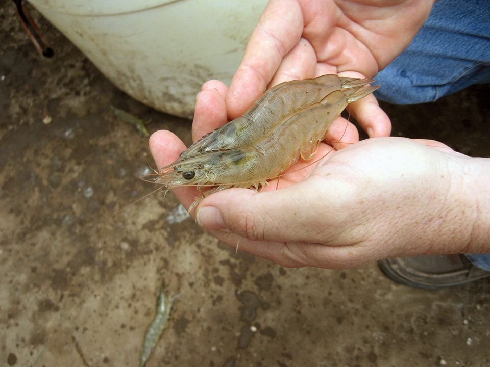
Trials measure the possible defense role of cuticle

Many experimental studies have demonstrated that shrimp and other decapods become infected with white spot syndrome virus (WSSV) through feeding on infected shrimp tissues. Many studies also reported that immersion and even cohabitation exposure readily allowed WSSV to cause infection, although older shrimp were sometimes reported to be less susceptible. These reports on WSSV helped to build the image that the virus is highly contagious.
On the other hand, several published studies showed that not all animals became infected through natural routes. Some researchers had to administer WSSV-infected tissues in more than one feeding, sometimes as long as seven days in order to infect the subject shrimp. More recent studies have faced serious difficulties in achieving infection, especially via the waterborne route.
It is important to note, however, that most of the early studies were performed with nonspecific pathogen-free (SPF) animals without knowing the doses of WSSV and without screening the inoculum for the presence of other pathogens.
Capacity to fight WSSV
Although few details are known about the structure and function of the cuticle of penaeid shrimp, it is well known that the cuticle changes dramatically over time. Also, it is often stated that the defense systems of Crustacea may be compromised at the time of molting.
On this basis, the authors designed a study to examine whether the physiological condition of shrimp in different molt stages influences their capacity to fight WSSV infection.
First, the virus was delivered intramuscularly, surpassing the cuticle, to estimate if any intrinsic difference in susceptibility to WSSV existed in shrimp in different stages of the molt cycle. Groups of SPF (Penaeus vannamei) in each of five major molt stages (A, B, C, D1 and D2) were injected intramuscularly with WSSV inoculum.
The results of this experiment showed that no significant difference in internal susceptibility to WSSV existed between shrimp in different molt stages. Hence, a series of trials was set up to mimic natural transmission through water and measure the possible defense role of cuticle.
Capacity to resist waterborne WSSV
The second experiment examined whether the stage in the molt cycle has an influence on the susceptibility of a shrimp to infection with WSSV. Shrimp in different molt stages were immersed in cell culture flasks containing WSSV inoculum for three hours. No food was given the first 12 hours after the immersion to avoid possible additional oral uptake of virus via the food. Animals were followed clinically, and WSSV infections were confirmed by indirect immunofluorescence.
Shrimp in molt stage A (early postmolt with thinnest cuticle) most commonly became infected (Table 1). Some in stage B and C were also infected, but no infection was seen in premolt stages. However, it was observed that some lesions in the cuticle were caused by the process of getting the shrimp inside the flasks. Most of the animals in which this was noted became infected. This observation gave rise to the hypothesis that the cuticle of shrimp presents a full barrier against WSSV, and that infection can only happen through wounds.
Alday-Sanz, Effects of immersion of P. vannamei at different molt stages,Table 1
| Weight | Molt Stage | Mortality (Hours After Inoculation) | Confirmed Infections |
|---|
Weight | Molt Stage | Mortality (Hours After Inoculation) | Confirmed Infections |
|---|---|---|---|
| 1 g | A B C D1 D2 | 1/5 (60) 0/5 0/5 0/5 0/5 | 1/5 0/5 0/5 0/5 0/5 |
| 4 g | A B C D1 D2 | 0/5 0/5 0/5 0/5 0/5 | 0/5 0/5 0/5 0/5 0/5 |
| 6 g | A B C D1 D2 | 3/5 (60, 60, 84) 0/5 0/5 0/5 0/5 | 3/5 0/5 0/5 0/5 0/5 |
| 11 g | A B C D1 D2 | 5/5 (48, 48, 48, 72, 72) 1/5 (120) 0/5 0/5 1/5 (During immersion) | 5/5 1/5 0/5 0/5 0/5 |
| 20 g | A B C D1 D2 | 5/5 (48, 60, 60, 60, 72) 2/5 (60, 60 1/5 (60) 0/5 0/5 | 5/5 2/5 1/5 0/5 0/5 |
Cuticle as defense
To test the hypothesis that intact cuticle is a defense barrier, a new experiment was designed. Plastic bags replaced the hard plastic flasks to hold shrimp, and a pleopod was removed from half of the shrimp at each molt stage just before the immersion challenge. The rest of the experimental protocol and conditions were kept identical to the previous one.
The most important finding was that WSSV infection rates were two to eight times higher in groups of artificially damaged shrimp than in the control shrimp. As in the previous experiment, the incidences of infection and mortality were clearly highest in shrimp immersed in WSSV inoculum during the postmolt stage (Table 2).
Alday-Sanz, Immersion of P. vannamei or P. monodon at different molt stages, Table 2
| Species | Weight | Appendage Removal | Molt Stage | Mortality (Hours After Inoculation) | Confirmed Infections |
|---|
Species | Weight | Appendage Removal | Molt Stage | Mortality (Hours After Inoculation) | Confirmed Infections |
|---|---|---|---|---|---|
| P. vannamei | 2 g | None (Control) | A B C D1 D2 | 2/5 (48, 72) 0/5 1/5 (72) 0/5 0/5 | 2/5 0/5 1/5 0/5 0/5 |
| P. vannamei | 1 pleopod | A B C D1 D2 | 3/5 (48, 60, 72) 3/5 (60, 60, 72) 1/5 (60) 0/5 0/5 | 3/5 3/5 1/5 0/5 0/5 |
|
| P. vannamei | 15 g | None (Control) | A B C D1 D2 | 1/5 (72) 2/5 (96, 96) 1/5 (120) 0/5 0/5 | 1/5 1/5 0/5 0/5 0/5 |
| P. vannamei | 1 pleopod | A B C D1 D2 | 5/5 (48, 72, 84, 84, 120) 2/5 (96, 96) 1/5 (120) 0/5 0/5 | 5/5 2/5 1/5 0/5 0/5 |
|
| P. monodon | 2g | None (Control) | A B C D1 D2 | 1/5 (72) 0/5 0/5 0/5 0/5 | 1/5 0/5 0/5 0/5 0/5 |
| P. monodon | 1 pleopod | A B C D1 D2 | 3/5 (48, 72, 72) 3/5 (72, 72, 84) 2/5 (72, 120) 0/5 0/5 | 3/5 3/5 2/5 0/5 0/5 |
|
| P. monodon | 15g | None (Control) | A B C D1 D2 | 2/5 (48, 72) 1/5 (48) 0/5 0/5 0/5 | 2/5 1/5 0/5 0/5 0/5 |
| P. monodon | 1 pleopod | A B C D1 D2 | 3/5 (48, 60, 72) 2/5 (48, 72) 3/5 (72, 84, 84) 0/5 0/5 | 3/5 2/5 3/5 0/5 0/5 |
Again, no infection was recorded in shrimp which were premolt at the time of exposure to waterborne virus, even with the infliction of a wound. While differences were seen in infection rates between ages in shrimp immersed in cell culture flasks, no such differences were recorded for shrimp of 2 to 15 grams inoculated inside plastic bags.
Molt stages and WSSV transmission
In shrimp, postmolt stages A and B are characterized by an epidermis in close contact with the entire still-thin cuticle. In the intermolt stage, the construction of the cuticle is finalized and the epidermis becomes inactive. The premolt phase starts as the epidermis retracts from the cuticle in stage D1 and begins formation of a new cuticle. In the final stage before the molt, D2, the newly forming cuticle and setae become visible. The proximity of susceptible epidermis cells to the cuticle in stages A and B may give WSSV a higher chance to access the host cells at the damaged site, allowing the establishment of the infection.
The authors’ studies suggested that if the cuticle of shrimp is fully intact, no WSSV infection will occur. Although damage to the cuticle appears to be key in WSSV infection from the water, the situation is surely more complex.
Natural outbreaks of WSSV in big pond populations are characterized by massive shrimp death within short periods of time. This suggests a fast and widespread transmission of WSSV.
It is still unclear whether the main route of entry of WSSV in natural outbreaks is vertical, via food and/or via water. These studies showed that cuticular damage can present a site of entry for waterborne virus, but at the same time indicated that high doses of the virus must be present in pond water and that large portions of the premolt population are not susceptible to waterborne infection. This indicated the main mode of WSSV infection might be through feeding on infected tissues, but perhaps microscopic lesions in the stomach or gill cuticle must be present to allow entry.
(Editor’s Note: This article was originally published in the July/August 2008 print edition of the Global Aquaculture Advocate.)
Now that you've finished reading the article ...
… we hope you’ll consider supporting our mission to document the evolution of the global aquaculture industry and share our vast network of contributors’ expansive knowledge every week.
By becoming a Global Seafood Alliance member, you’re ensuring that all of the pre-competitive work we do through member benefits, resources and events can continue. Individual membership costs just $50 a year. GSA individual and corporate members receive complimentary access to a series of GOAL virtual events beginning in April. Join now.
Not a GSA member? Join us.
Authors
-
Victoria Alday-Sanz, Ph.D.
Aquatic Animal Health Consultant
Gran Via 658, 4-1
08010 Barcelona, Spain -
M. Corteel
Laboratory of Virology
Faculty of Veterinary Medicine
Ghent University
Merelbeke, Belgium -
J.J. Dantas-Lima
Laboratory of Virology
Faculty of Veterinary Medicine
Ghent University
Merelbeke, Belgium -
M.B. Pensaert
Laboratory of Virology
Faculty of Veterinary Medicine
Ghent University
Merelbeke, Belgium -
H.J. Nauwynck
Laboratory of Virology
Faculty of Veterinary Medicine
Ghent University
Merelbeke, Belgium -
M. Wille
Laboratory of Aquaculture & Artemia Reference Center
Faculty of Bioscience Engineering
Ghent University
Ghent, Belgium -
Dr. P. Sorgeloos
Laboratory of Aquaculture & Artemia Reference Center
Faculty of Bioscience Engineering
Ghent University
Ghent, Belgium
Tagged With
Related Posts

Health & Welfare
A holistic management approach to EMS
Early Mortality Syndrome has devastated farmed shrimp in Asia and Latin America. With better understanding of the pathogen and the development and improvement of novel strategies, shrimp farmers are now able to better manage the disease.

Health & Welfare
A study of Zoea-2 Syndrome in hatcheries in India, part 2
Indian shrimp hatcheries have experienced larval mortality in the zoea-2 stage, with molt deterioration and resulting in heavy mortality. Authors considered biotic and abiotic factors. Part 2 describes results of their study.

Health & Welfare
A study of Zoea-2 Syndrome in hatcheries in India, part 3
In this third and final part, authors present recommendations to help reduce the incidence of Zoea-2 Syndrome, which is not caused by any known infectious agents in P. vannamei hatcheries in India.

Health & Welfare
AHPN inferences based on behavior of vibrio bacteria
Vibrio parahaemolyticus, a strain of which is the cause of acute hepatopancreatic necrosis (AHPN), has both virulent and benign strains. This strain colonizes the stomachs of shrimp by the formation of a biofilm, which protects it from antibiotics and other potential treatments.


