Hatchery management, pathogen detection and results analysis

Editor’s note: This is part 2 of a series. Click here to read part 1.
During the study, nine hatcheries were affected by the Zoea-2 Syndrome and six hatcheries – including two nauplii rearing centers in Andhra Pradesh – were not affected and had healthy larval production cycles. All hatcheries used breeding populations of imported, specific pathogen free (SPF) Pacific White shrimp (Penaeus vannamei) for seedstock production. Broodstock animals were fed fresh polychaetes, squid, oysters and an artificial pelleted diet.
In the spawning tanks, ethylenediaminetetraacetic acid (EDTA) was used as a chelating agent for heavy metals, and the fungicide treflan was also used. After each spawning, the eggs were washed with formaldehyde (100 ppm for 30 seconds), with iodine (50-100 ppm for 1 minute) and with seawater before stocking. (Table 1).
Kumar, Zoea-2, Table 1
| Hatchery | Location | Zoea-2 Syndrome | Egg washing | Probiotics used | Antibiotics used | Algae used | Temperature | pH | DO | Salinity | Alkalinity |
|---|
Hatchery | Location | Zoea-2 Syndrome | Egg washing | Probiotics used | Antibiotics used | Algae used | Temperature | pH | DO | Salinity | Alkalinity |
|---|---|---|---|---|---|---|---|---|---|---|---|
| A | Marakanam, TN | Affected | Formaldehyde, iodine | B. subtilis, B. litcheniforms | Oxytetracycline | Chaetoceros | 30 | 7.1 | 5 | 34 | 145 |
| B | Marakanam, TN | Affected | Formaldehyde, iodine | Bacillus sp. | Oxytetracycline | Skeletonema, Thalassiosira | 29 | 7.7 | 4 | 30 | 120 |
| C | Marakanam, TN | Normal | Formaldehyde, iodine | Bacillus sp. Y Pseudomonas | Oxytetracycline, Erythromycin | Chaetoceros, Skeletonema | 29 | 8.1 | 5 | 31 | 115 |
| D | Marakanam, TN | Affected | Formaldehyde, iodine, treflan | Bacillus sp., Lactobacillus sp. | Oxytetracycline | Chaetoceros, Thalassiosira | 30 | 8 | 5 | 31 | 110 |
| E | Marakanam, TN | Normal | Formaldehyde, iodine | Bacillus sp. | None | Chaetoceros, Skeletonema | 30 | 7.5 | 4 | 30 | 135 |
| F | Marakanam, TN | Normal | Formaldehyde, iodine | B. subtilis y Rhodopseudomonas | Oxytetracycline | Chaetoceros, Thalassiosira | 29 | 7 | 6 | 31 | 120 |
| G | Nellore, AP | Affected | Formaldehyde, iodine | B. subtilis, B. Pumilis, B. Megaterium | Oxytetracycline, Erythromycin | Chaetoceros, Thalassiosira | 28 | 7.5 | 5 | 30 | 115 |
| H | Nellore, AP | Affected | Formaldehyde, iodine, treflan | Bacillus sp. | Oxytetracycline | Chaetoceros, Thalassiosira | 29 | 7.3 | 4 | 31 | 140 |
| I | Nellore, AP | Normal | Formaldehyde, iodine | Bacillus sp., y Streptococcus sp. | Oxytetracycline, Erythromycin | Chaetoceros | 29 | 8 | 4 | 31 | 125 |
| J | Ongole, AP | Affected | Formaldehyde, iodine | Bacillus sp., Lactobacillus sp. | Oxytetracycline | Chaetoceros, Thalassiosira | 30 | 7.8 | 4 | 30 | 110 |
| Normal | Formaldehyde, iodine | Bacillus sp., Lactobacillus sp. | Oxytetracycline | – | 29 | 8 | 5 | 30 | 130 | ||
| K | Ongole, AP | Affected | Formaldehyde, iodine, treflan | Bacillus sp., Levadura | None | Chaetoceros, Thalassiosira | 28 | 7.5 | 6 | 30 | 120 |
| L | Ongole, AP (NCR) | Normal | Formaldehyde, iodine, treflan | Lactobacillus sp. | Oxytetracycline | Skeletonema, Chaetoceros, Thalassiosira | 30 | 8 | 5 | 31 | 120 |
| M | Kakinada, AP (NCR) | Affected | Formaldehyde, iodine | Bacillus sp. | None | Chaetoceros, Thalassiosira | 29 | 7.5 | 6 | 29 | 115 |
| N | Kakinada, AP | Affected | Formaldehyde, iodine, treflan | B. punctatuts, B. Subtilis | Oxytetracycline | Chaetoceros, Thalassiosira | 31 | 8 | 6 | 30 | 135 |
| O | Kakinada, AP | Affected | Formaldehyde, iodine | Bacillus sp. | Oxytetracycline | Chaetoceros, Thalassiosira | 29 | 7.3 | 4 | 29 | 130 |
Next, the nauplii in the stage N-VI were stocked in larval rearing units and reared to PL. This production cycle continued with daily spawnings, followed by daily stocking of nauplii (3 to 10 days) in the next larval rearing tanks. A continuous stocking of nauplii was observed for more than four days in nine hatcheries affected by Zoea-2 Syndrome and in one normal, unaffected hatchery (C).
Water quality parameters (pH, salinity and alkalinity) in all hatcheries were within normal ranges (Table 1) and did not seem to influence the occurrence of Zoea-2 Syndrome. At all the hatcheries, the seawater treatment protocol involved sedimentation, chlorination, de-chlorination and filtration (sand filter, activated charcoal and cartridge filters) followed by UV filtration and ozonation. Oxytetracycline and probiotic formulations containing Bacillus sp., Streptococcus sp. and Lactobacillus sp. were used in the hatcheries. The microalgae Skeletonema, Chaetoceros and Thalassiosira were used as feed during the zoeal stages.
Kumar, Zoea-2, Table 2
| Hatchery | Zoea-2 Syndrome | Stocking | Deficiency in unit separation | Deficiency of adequate disinfection | Swimming activity | Phototaxis | Empty guts | Sudden feeding stoppage | Peristaltic gut movement stoppage | Gut inflammation | Fecal strands | White balls | Bacteria isolated |
|---|
Hatchery | Zoea-2 Syndrome | Stocking | Deficiency in unit separation | Deficiency of adequate disinfection | Swimming activity | Phototaxis | Empty guts | Sudden feeding stoppage | Peristaltic gut movement stoppage | Gut inflammation | Fecal strands | White balls | Bacteria isolated |
|---|---|---|---|---|---|---|---|---|---|---|---|---|---|
| A | Affected | Yes | No | Yes | No | No | Yes | Yes | Yes | Yes | No | No | V. alginolyticus, V. mimicus |
| B | Affected | Yes | Yes | Yes | No | No | Yes | Yes | Yes | Yes | No | Yes | V. alginolyticus |
| C | Normal | Yes | No | No | Yes | Yes | No | No | No | No | Yes | No | V. alginolyticus, V. campbelli |
| D | Affected | Yes | No | Yes | No | No | Yes | Yes | Yes | Yes | No | Yes | V. alginolyticus, V. mimicus |
| E | Normal | No | Yes | No | Yes | Yes | No | No | No | No | Yes | No | V. alginolyticus |
| F | Normal | No | No | Yes | Yes | Yes | No | No | No | No | Yes | No | V. metschnikonii, V. mimicus |
| G | Affected | No | Yes | Yes | No | No | Yes | Yes | Yes | Yes | No | No | V. alginolyticus |
| H | Affected | Yes | Yes | Yes | No | No | Yes | Yes | Yes | Yes | No | Yes | V. alginolyticus, V. vulnificus |
| I | Normal | No | Yes | No | Yes | Yes | No | No | No | No | Yes | No | V. alginolyticus, V. proteolyticus |
| J | Affected | Yes | No | No | No | No | Yes | No | Yes | Yes | No | Yes | V. alginolyticus, V. mimicus, V. furnissi |
| Normal | No | No | No | Yes | Yes | No | No | No | No | Yes | No | V. mytili, V. mimicus | |
| K | Affected | Yes | Yes | Yes | No | No | Yes | Yes | Yes | Yes | No | Yes | V. alginolyticus, V. mimicus |
| L | Normal | No | No | No | Yes | Yes | No | No | No | No | Yes | No | V. cincinatensis |
| M | Affected | No | No | Yes | Yes | Yes | No | No | No | No | Yes | No | V. cincinatensis, V. mimicus |
| N | Affected | Yes | Yes | Yes | No | No | Yes | Yes | Yes | Yes | No | Yes | V. cincinatensis, V. vulnificus |
| O | Affected | Yes | No | Yes | No | No | Yes | Yes | Yes | Yes | No | No | V. alginolyticus, V. mimicus |
After each larval production cycle, the tanks and equipment were disinfected with hypochlorite solution (20 to 30 ppm active ingredient); the water pipes were disinfected by filling with disinfectant solution (chlorine, 500 ppm; potassium permanganate (KMnO4), 20 ppm; formaldehyde, 200 ppm; muriatic acid, 10 percent), and the air pipes were disinfected by fumigation with formaldehyde (200 ppm).

Two hatcheries (B, G) had no separate algae culture units, and in three hatcheries (E, H, N), the same workers were allowed to rotate to/from different units. In one hatchery (G), the same air blower was shared by two larval breeding units. Lack of disinfection was also observed during and between the larval production cycles in eight hatcheries affected by the Zoea-2 Syndrome and in two normal, unaffected hatcheries (Table 2).
A batch of nauplii produced in a hatchery (J) was stocked on the fourth day in larval rearing tanks in a larviculture unit that had previous batches of affected larvae. Some of the nauplii from the same batch were stocked in another unit in a different section in the same hatchery. The nauplii stored in the section with older, affected larvae developed the Zoea-2 Syndrome and did not metamorphose into mysis and PL. The same nauplii stocked in the other section in the same hatchery had no anomalies and metamorphosed into healthy mysis and PL.
In another case, the nauplii breeding center (M) produced PLs without any problems, while the same batch of nauplii stocked in the hatchery (K) of the producers (which already had Zoea-2 Syndrome) was affected by the syndrome. The incidences of the zoea syndrome were low in the hatcheries without maturation units (hatcheries L, M: these data are not presented).
Bacteriology
A total of 29 dominant vibrios were isolated from the 15 hatcheries. Of the nine affected hatcheries, Vibrio alginolyticus was found predominantly in eight, followed by V. mimicus in five and V. vulnificus in two hatcheries. In six hatcheries with no Zoea-2 Syndrome, V. alginolyticus and V. mimicus were predominant in three, followed by V. cincinatensis in two. In one hatchery (J), V. alginolyticus was predominant, followed by V. mimicus and V. furnissi.
Detection of viral pathogens
To determine if the presence of any known viral agent may have had a role in causing the Zoea-2 Syndrome, all the samples of larvae collected from the different hatcheries were screened for the OIE-registered viral pathogens, including WSSV, MBV, IHHNV, YHV, IMNV, TSV and CMNV. All larval samples collected from the hatcheries affected by the Zoea-2 Syndrome were negative for these DNA and RNA viruses.
Light microscopy exam

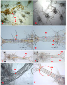
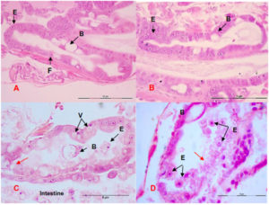
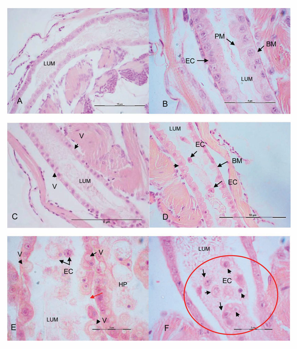
Recently collected live and healthy animals, as well as affected animals (after 36 to 48 hours of stage zoea I), were observed under a light microscope. Normal, unaffected zoea showed an active peristaltic movement of the intestine filled with food and long fecal strands projected from the anus (Fig. 1A, C, E). The affected zoea were less active and exhibited an almost empty intestine, with weak peristaltic movement and without fecal strands. The intestinal lumen showed inflammation (Fig. 1B, D, F, G and H). In histology, the hepatopancreas of normal zoea had intact tubules with developing B, F, and E cells (Fig. 2A, B), while the hepatopancreas of affected zoea showed severe necrosis and detachment of epithelial cells from the basal membrane of the hepatopancreatic tubule epithelium (Fig. 2C, D). The longitudinal histological sections of the intestine showed hypertrophy (Fig. 3E, F), vacuolization in columnar epithelial cells (Fig. 3C, D, E), peritrophic membrane disintegration (Fig. 3D, E) and tearing / desquamation of the epithelial cells of the basal membrane epithelium in the lumen of the intestine (Fig. 3D, E, F), compared to normal zoea that did not exhibit these systemic abnormalities (Fig. 3A, B).

Electron transmission microscope
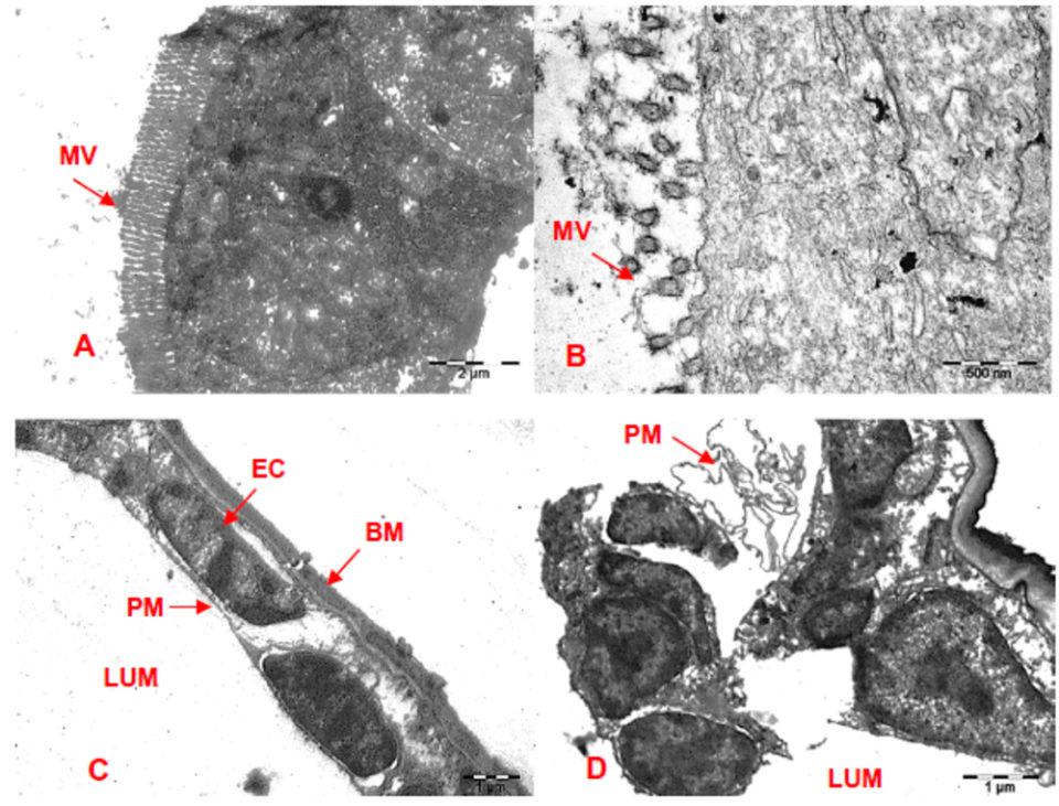
Ultrastructural studies revealed detachment of microvilli from the epithelial cells in hepatopancreatic tubules (Fig. 4B) compared to normal hepatopancreas with intact microvilli (Fig. 4A). Similarly, intestines in affected zoea showed disintegration and detachment of the peritrophic membrane; and also necrosis, desquamation and epithelial cell detachment from the basal membrane in the intestinal epithelium (Fig. 4D) compared to the normal intestine with an intact epithelium (Fig. 4C). No viral particles were observed in the ultramicroscopic sections.

Statistical and epidemiological analysis
During the study it was observed that the incidences of zoea syndrome were more common in hatcheries with prolonged larval production cycles with continuous storage of nauplii for more than four days in the same larval breeding unit; with lack of adequate disinfection between cycles; and with broodstock animals that did not have a separate maturation unit, separate larval rearing and algae culture units, and different workers and equipment/implements for different units (Table 2).
To find an association between the incidence factors of the zoea-2 syndrome and the exposure of hatcheries mentioned above, the probability coefficient (p-value) was calculated. The p-value for the two hatchery factors – the storage of nauplii for more than four days in the same unit and the lack of adequate disinfection – was >1. Although the p-value with respect to the lack of separate units was >1, its confidence interval (CI 0.38-25.5) goes through 1. The storage of nauplii for more than four days in the same unit and the lack of adequate disinfection were both significantly associated with the increased incidence of zoea-2 syndrome, while the lack of separate units had no significant association (Table 3).
Kumar, Zoea-2, Table 3
| Hatchery factors | Possibility index | Lower confidence interval (95%) | Upper confidence interval (95%) | P value |
|---|
Hatchery factors | Possibility index | Lower confidence interval (95%) | Upper confidence interval (95%) | P value |
|---|---|---|---|---|
| Nauplii storage, over 4 days in same unit | 48 | 2.4697 | 932.9003 | 0.008 |
| Deficiency in unit separation | 3,125 | 0.382 | 25.5669 | 0.35 |
| Deficiency in appropriate disinfection | 20 | 1.4161 | 282.4627 | 0.03 |
Authors
-
Sathish Kumar
ICAR-Central Institute of Brackishwater Aquaculture
#75 Santhome High Road, Raja Annamalai Puram
Chennai 600028, India -
Vidya
ICAR-Central Institute of Brackishwater Aquaculture
#75 Santhome High Road, Raja Annamalai Puram
Chennai 600028, India -
Dr. Sujeet Kumar
ICAR-Central Institute of Brackishwater Aquaculture
#75 Santhome High Road, Raja Annamalai Puram
Chennai 600028, India -
Dr. S.V. Alavandi
ICAR-Central Institute of Brackishwater Aquaculture
#75 Santhome High Road, Raja Annamalai Puram
Chennai 600028, India -
Dr. K.K. Vijayan
ICAR-Central Institute of Brackishwater Aquaculture
#75 Santhome High Road, Raja Annamalai Puram
Chennai 600028, India
Tagged With
Related Posts

Health & Welfare
A study of Zoea-2 Syndrome in hatcheries in India, part 1
Indian shrimp hatcheries have experienced larval mortality in the zoea-2 stage, with molt deterioration and resulting in heavy mortality. Authors investigated the problem holistically.

Health & Welfare
How good are your shrimp postlarvae?
Stocking the best quality shrimp postlarvae, healthy and free of pathogens, is a critical management step with significant effects on the production and profitability of a shrimp farm.

Innovation & Investment
Shrimp farming in China: Lessons from its developmental history
Fenneropenaeus chinensis was the most important farmed shrimp species in China until 1995. Lessons learned from its development made China a pioneer, especially in shrimp larval production. Shrimp farmers must enhance their understanding of the interactions of farming activities with their ecosystems.

Health & Welfare
Vibrio control in shrimp farming: Part 1
Control of Vibrio bacteria should focus on minimizing overall bacterial loads and the potential for horizontal transmission. The challenge for hatchery managers is identifying gaps in biosecurity and plugging them without creating niches for other potential pathogens.


